8HMA

Cryo-EM structure of human high-voltage activated L-type calcium channel CaV1.2 in complex with tetrandrine (TET)


Experimental Data Snapshot

  • Method: ELECTRON MICROSCOPY
  • Resolution: 3.40 Å
  • Aggregation State: PARTICLE 
  • Reconstruction Method: SINGLE PARTICLE 

wwPDB Validation   3D Report Full Report


This is version 1.0 of the entry. See complete history


Literature

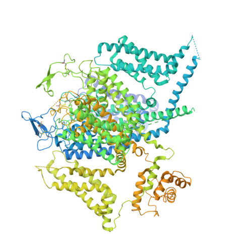
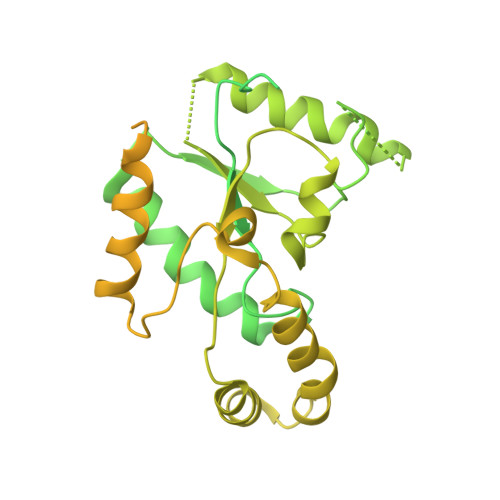
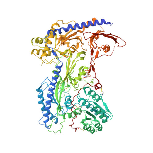
Structural bases of inhibitory mechanism of Ca V 1.2 channel inhibitors.

Wei, Y.Yu, Z.Wang, L.Li, X.Li, N.Bai, Q.Wang, Y.Li, R.Meng, Y.Xu, H.Wang, X.Dong, Y.Huang, Z.Zhang, X.C.Zhao, Y.

(2024) Nat Commun 15: 2772-2772

  • DOI: https://doi.org/10.1038/s41467-024-47116-8
  • Primary Citation of Related Structures:  
    8HLP, 8HMA, 8HMB

  • PubMed Abstract: 

    The voltage-gated calcium channel Ca V 1.2 is essential for cardiac and vessel smooth muscle contractility and brain function. Accumulating evidence demonstrates that malfunctions of Ca V 1.2 are involved in brain and heart diseases. Pharmacological inhibition of Ca V 1.2 is therefore of therapeutic value. Here, we report cryo-EM structures of Ca V 1.2 in the absence or presence of the antirheumatic drug tetrandrine or antihypertensive drug benidipine. Tetrandrine acts as a pore blocker in a pocket composed of S6 II , S6 III , and S6 IV helices and forms extensive hydrophobic interactions with Ca V 1.2. Our structure elucidates that benidipine is located in the D III -D IV fenestration site. Its hydrophobic sidechain, phenylpiperidine, is positioned at the exterior of the pore domain and cradled within a hydrophobic pocket formed by S5 DIII , S6 DIII , and S6 DIV helices, providing additional interactions to exert inhibitory effects on both L-type and T-type voltage gated calcium channels. These findings provide the structural foundation for the rational design and optimization of therapeutic inhibitors of voltage-gated calcium channels.


  • Organizational Affiliation

    Key Laboratory of Biomacromolecules (CAS), National Laboratory of Biomacromolecules, CAS Center for Excellence in Biomacromolecules, Institute of Biophysics, Chinese Academy of Sciences, Beijing, 100101, China.


Macromolecules
Find similar proteins by:  (by identity cutoff)  |  3D Structure
Entity ID: 1
MoleculeChains Sequence LengthOrganismDetailsImage
Voltage-dependent L-type calcium channel subunit alphaA [auth E]2,173Homo sapiensMutation(s): 0 
Gene Names: CACNA1C
UniProt & NIH Common Fund Data Resources
Find proteins for A0A0A0MSA1 (Homo sapiens)
Explore A0A0A0MSA1 
Go to UniProtKB:  A0A0A0MSA1
GTEx:  ENSG00000151067 
Entity Groups  
Sequence Clusters30% Identity50% Identity70% Identity90% Identity95% Identity100% Identity
UniProt GroupA0A0A0MSA1
Sequence Annotations
Expand
  • Reference Sequence
Find similar proteins by:  (by identity cutoff)  |  3D Structure
Entity ID: 2
MoleculeChains Sequence LengthOrganismDetailsImage
Isoform 2c of Voltage-dependent L-type calcium channel subunit beta-2B [auth H]632Homo sapiensMutation(s): 0 
Gene Names: CACNB2CACNLB2MYSB
UniProt & NIH Common Fund Data Resources
Find proteins for Q08289 (Homo sapiens)
Explore Q08289 
Go to UniProtKB:  Q08289
PHAROS:  Q08289
GTEx:  ENSG00000165995 
Entity Groups  
Sequence Clusters30% Identity50% Identity70% Identity90% Identity95% Identity100% Identity
UniProt GroupQ08289
Sequence Annotations
Expand
  • Reference Sequence
Find similar proteins by:  (by identity cutoff)  |  3D Structure
Entity ID: 3
MoleculeChains Sequence LengthOrganismDetailsImage
Voltage-dependent calcium channel subunit alpha-2/delta-1C [auth F]1,071Homo sapiensMutation(s): 0 
Gene Names: CACNA2D1CACNL2ACCHL2AMHS3
UniProt & NIH Common Fund Data Resources
Find proteins for P54289 (Homo sapiens)
Explore P54289 
Go to UniProtKB:  P54289
PHAROS:  P54289
GTEx:  ENSG00000153956 
Entity Groups  
Sequence Clusters30% Identity50% Identity70% Identity90% Identity95% Identity100% Identity
UniProt GroupP54289
Sequence Annotations
Expand
  • Reference Sequence
Small Molecules
Ligands 6 Unique
IDChains Name / Formula / InChI Key2D Diagram3D Interactions
3PE
Query on 3PE

Download Ideal Coordinates CCD File 
E,
F [auth E],
H [auth E]
1,2-Distearoyl-sn-glycerophosphoethanolamine
C41 H82 N O8 P
LVNGJLRDBYCPGB-LDLOPFEMSA-N
A1AC9 (Subject of Investigation/LOI)
Query on A1AC9

Download Ideal Coordinates CCD File 
G [auth E]6,6',7,12-tetramethoxy-2,2'-dimethyl-1beta-3,4-didehydroberbaman
C38 H40 N2 O6
OUHWUXGBWHCKDH-UFXYQILXSA-N
R16
Query on R16

Download Ideal Coordinates CCD File 
D [auth E]HEXADECANE
C16 H34
DCAYPVUWAIABOU-UHFFFAOYSA-N
NAG
Query on NAG

Download Ideal Coordinates CCD File 
K [auth F]
L [auth F]
M [auth F]
N [auth F]
P [auth F]
K [auth F],
L [auth F],
M [auth F],
N [auth F],
P [auth F],
Q [auth F],
R [auth F],
S [auth F],
T [auth F],
U [auth F],
V [auth F]
2-acetamido-2-deoxy-beta-D-glucopyranose
C8 H15 N O6
OVRNDRQMDRJTHS-FMDGEEDCSA-N
BMA
Query on BMA

Download Ideal Coordinates CCD File 
O [auth F]beta-D-mannopyranose
C6 H12 O6
WQZGKKKJIJFFOK-RWOPYEJCSA-N
CA
Query on CA

Download Ideal Coordinates CCD File 
I [auth E],
J [auth E]
CALCIUM ION
Ca
BHPQYMZQTOCNFJ-UHFFFAOYSA-N
Experimental Data & Validation

Experimental Data

  • Method: ELECTRON MICROSCOPY
  • Resolution: 3.40 Å
  • Aggregation State: PARTICLE 
  • Reconstruction Method: SINGLE PARTICLE 

Structure Validation

View Full Validation Report


Entry History & Funding Information

Deposition Data


Funding OrganizationLocationGrant Number
Chinese Academy of SciencesChina--

Revision History  (Full details and data files)

  • Version 1.0: 2024-04-24
    Type: Initial release