


Crystal structure of CotB2 variant V80L in complex with alendronate
Dimos, N., Himpich, S., Driller, R., Loll, B.To be published.
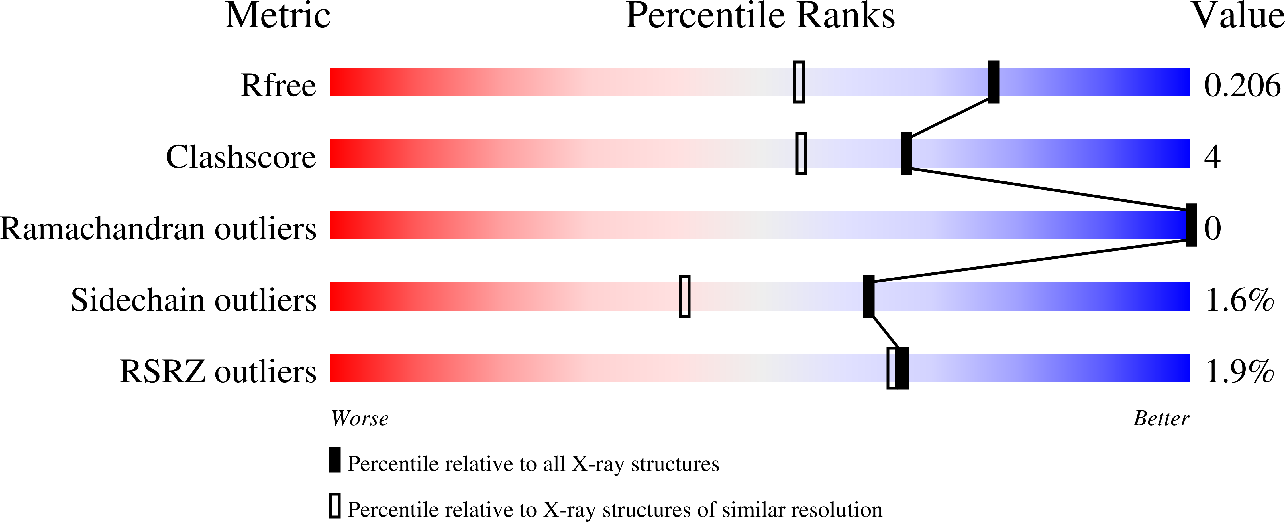
Experimental Data Snapshot
Entity ID: 1 | |||||
|---|---|---|---|---|---|
| Molecule | Chains | Sequence Length | Organism | Details | Image |
| Cyclooctat-9-en-7-ol synthase | 318 | Streptomyces melanosporofaciens | Mutation(s): 1 Gene Names: CotB2 | 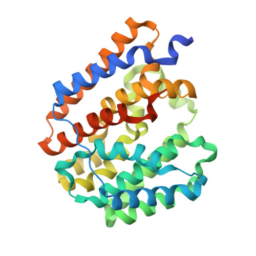 | |
UniProt | |||||
Find proteins for C9K1X5 (Streptomyces melanosporofaciens) Explore C9K1X5 Go to UniProtKB: C9K1X5 | |||||
Entity Groups | |||||
| Sequence Clusters | 30% Identity50% Identity70% Identity90% Identity95% Identity100% Identity | ||||
| UniProt Group | C9K1X5 | ||||
Sequence AnnotationsExpand | |||||
| |||||
| Ligands 7 Unique | |||||
|---|---|---|---|---|---|
| ID | Chains | Name / Formula / InChI Key | 2D Diagram | 3D Interactions | |
| AHD (Subject of Investigation/LOI) Query on AHD | C [auth A], S [auth B] | 4-AMINO-1-HYDROXYBUTANE-1,1-DIYLDIPHOSPHONATE C4 H9 N O7 P2 OGSPWJRAVKPPFI-UHFFFAOYSA-J |  | ||
| PG0 Query on PG0 | FA [auth B], Q [auth A] | 2-(2-METHOXYETHOXY)ETHANOL C5 H12 O3 SBASXUCJHJRPEV-UHFFFAOYSA-N |  | ||
| MPD Query on MPD | BA [auth B], GA [auth B], HA [auth B], J [auth A] | (4S)-2-METHYL-2,4-PENTANEDIOL C6 H14 O2 SVTBMSDMJJWYQN-YFKPBYRVSA-N |  | ||
| EDO Query on EDO | CA [auth B] EA [auth B] G [auth A] H [auth A] I [auth A] | 1,2-ETHANEDIOL C2 H6 O2 LYCAIKOWRPUZTN-UHFFFAOYSA-N |  | ||
| ACT Query on ACT | AA [auth B], DA [auth B], L [auth A], O [auth A], P [auth A] | ACETATE ION C2 H3 O2 QTBSBXVTEAMEQO-UHFFFAOYSA-M |  | ||
| CL Query on CL | X [auth B] | CHLORIDE ION Cl VEXZGXHMUGYJMC-UHFFFAOYSA-M |  | ||
| MG Query on MG | D [auth A] E [auth A] F [auth A] T [auth B] U [auth B] | MAGNESIUM ION Mg JLVVSXFLKOJNIY-UHFFFAOYSA-N |  | ||
| Length ( Å ) | Angle ( ˚ ) |
|---|---|
| a = 61.173 | α = 90 |
| b = 98.514 | β = 90 |
| c = 107.417 | γ = 90 |
| Software Name | Purpose |
|---|---|
| PHENIX | refinement |
| XDS | data reduction |
| XSCALE | data scaling |
| PHASER | phasing |
| Funding Organization | Location | Grant Number |
|---|---|---|
| German-Israeli Foundation for Research and Development | Germany | I-85-302.5-2019 |
RCSB PDB (citation) is hosted by
RCSB PDB is a member of the

